You have no items in your shopping cart.
Synonym(s):
Coomassie dye binding protein assay, Coomassie dye binding protein assay, Protein dye reagent, Protein dye reagent
Introduction
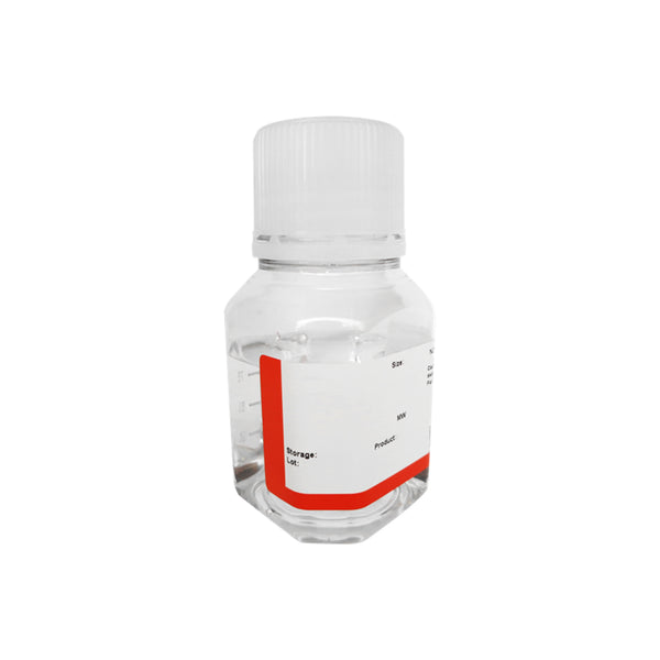
Bradford method utilizes Coomassie Brillant Blue G-250 dye binding to an unknown protein and forming a complex which can be detected spectophotometrically at 595 nm.
Key Features
-
Rapid and easy-to-use: Entire assay (10–20 samples) takes only ~10 minutes (excluding sample prep)
-
High sensitivity: Detects as little as 0.2 µg of protein in 1–20 µL
-
Excellent linearity: 10–150 µg/mL linear detection range
-
Versatile: Compatible with both cuvette-based and 96-well plate assays
-
Cost-effective alternative to more expensive protein quantification kits
Application
-
Quantitative measurement of total protein concentration in biological or biochemical samples
-
Protein estimation in cell lysates, column fractions, purified protein preparations, etc.
Assay Protocol Overview
Tube-Based Protocol
-
Prepare BSA standard curve (125–0 µg/mL) via serial dilution
-
Add 100 µL of each standard or sample to test tubes
-
Add 1 mL Bradford Reagent, mix and incubate at RT for 10 min
-
Measure absorbance at 595 nm
96-Well Plate Protocol
-
Add 20 µL of each sample or standard into wells
-
Add 200 µL of Bradford Reagent
-
Shake plate 30 sec, incubate 10 min at RT
-
Read at 595 nm with a microplate reader
Procedures
Protocols for Measurements in Test Tubes A
Preparation of BSA Standards and Sample Dilutions
-
Label eight test tubes from #1 to #8.
-
Prepare serial dilutions of the BSA standard as follows:
-
Tube #1: Add 20 µL of BSA standard (5 mg/mL) to 780 µL of PBS.
-
Tube #2: Mix 100 µL of PBS with 400 µL from Tube #1.
-
Tube #3: Mix 100 µL of PBS with 400 µL from Tube #2.
-
Tube #4: Mix 120 µL of PBS with 360 µL from Tube #3.
-
Tube #5: Mix 120 µL of PBS with 240 µL from Tube #4.
-
Tube #6: Mix 150 µL of PBS with 150 µL from Tube #5.
-
Tube #7: Mix 100 µL of PBS with 100 µL from Tube #6.
-
Tube #8: Add 100 µL of PBS only (blank control).
This is a serial dilution of the BSA standard.
| 1 | 2 | 3 | 4 | 5 | 6 | 7 | 8 | |
| BSA final concentration (μg/ml) |
125 | 100 | 80 | 60 | 40 | 20 | 10 | 0 |
Dilute the sample of interest to a desired concentration (10–125 μg/mL) and note the dilution factor (X).
Label Tubes #9 to #11
-
In Test Tube #9, transfer 20 µL of the diluted protein sample.
-
In Tube #10, transfer 20 µL of the diluted protein sample at a different dilution factor.
-
In Tube #11, transfer 20 µL of PBS only.
B. Measure the concentration of BSA standards and the sample of interest.
-
Transfer standards #1 to #8 into designated wells of a 96-well plate in duplicate to ensure reliable results.
-
Add 200 µl of Bradford Reagent to each well.
-
Shake the plate for 30 seconds to mix thoroughly.
-
Incubate at room temperature (RT) for 10 minutes.
-
Measure absorbance at 595 nm using a microplate reader.
-
Subtract the average absorbance of blank replicates from all standard and sample readings.
-
Plot a standard curve: average blank-corrected absorbance vs. BSA concentration (µg/ml).
-
Determine the protein concentration of your diluted samples using the standard curve, then calculate the original concentration by applying the dilution factor.
Notes
-
Mix reagent thoroughly before use; bring to room temperature
-
Avoid detergents (use BCA assay instead if sample contains detergents)
(SK3021, SK3051) -
For best results, prepare a standard curve using the same protein as the sample
-
Duplicate measurements and fresh standard curves are strongly recommended
Comparable To:
-
Sigma-Aldrich Bradford Reagent (B6916), but more economical
For Research Use Only. Not for human or animal therapeutic use.
Documents
Disclaimer: For laboratory research use only.
When can I expect my order to ship?
Most orders are filled and shipped within 2-3 business days from the time they are received.
Our standard shipping usually take 2-5 days.
We also provide express shippping for time-sensitive deliveries.
Email contact@biofargo.com if you have any requirements.


